Toshinori Kato

M.D., Ph.D., specializes in theoretical life science,
brain functional physiology, brain imaging, and photic
brain function measurement.
Professional profiles
At age 14, inspired by athletic training, I realized the important role of the brain, and determined to go to a medical school.
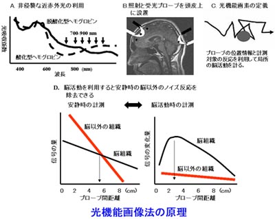
In 1991 at the National Center of Neurology and Psychiatry in Japan I discovered a principle to visualize function-related capillary response in brain areas engaged in different tasks by lighting from the scalp - photon functional imaging or Near-infrared spectroscopy (NIRS)-Imaging (fig.1).
This revolutionary idea was inspired by my experience researching child development by MR-spectroscopy with surface coil, brain MRI, and single-photon emission computed tomography (SPECT), and by medical practice in pediatrics and neonatology.
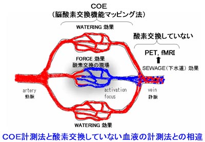
Photon functional pixels were defined by a different measuring principle from photon-CT (pCT) invented by Jobsis in 1977, and made technological progress from brain oxygenation monitoring to cerebral oxygen exchange imaging at bedside (fig.2).

This essential technology for sampling photon functional pixels led to a new brain function measuring device from Japan, and became a metabolism mapping system at bedside such as mapping blood flow metabolism and muscular metabolism. Now they are used worldwide.
I continued developing the principle to illuminate brain functions toward perfection.
In the same year I also invented a basic principle of cerebral white matter visualization by magnetic resonance imaging (MRI) and was acknowledged by Dr. Paul Christian Lauterbur, a winner of the Nobel Prize in Medicine in 2003 - a pioneer of magnetic resonance imaging.
From 1995 under Dr. Ogawa's guidance who was awarded The Japan Prize for research on fMRI with T2 weighted images. I was employed at the University of Minnesota Center for Magnetic Resonance Research.
My major research objective was to study higher brain functions such as the physiological activity of the hippocampus involving human memory function that was crucial for understanding of Alzheimer's disease and aging.
After six years of intensive research, I discovered a critical difference between the principle of photon functional imaging and measurement principles of fMRI, and led "hemo-dynamic-bridge theory" that connected NIRS-imaging and fMRI (fig.3).
The crucial difference between them was that fMRI targeted cerebral response in the veins, while photon functional imaging visualized cerebral capillary response.
This cerebral response in the vein was in fact not the response of the brain but a response outside of the brain.
Therefore, I realized that nobody had measured oxygen consumption in the cerebral blood flow for 110 years.
In 2001 while I was in the United States, I dissolved an imperfect dogma about neurons and brain blood flow that had been believed since the 19th century when Sir Charles Scott Sherrington, who won the Nobel Prize in medicine for discovering synapses and others proposed, and discovered “oxygen exchange wave equation” which described a life phenomenon with its imaginary number.
This way life phenomenon was formulated as a micro-system.
It had been proven by measurable value that living organisms which depended on oxygen exchange had imaginary dimension.
At this point, the progress of the principle of photon functional pixel imaging that illuminates brain function completed as Cerebral mapping of Oxygen Exchange (COE) that measured oxygen exchange systems.
This "oxygen exchange wave equation" achieved quantitative measurement of mental activity in the brain. It took cerebral oxygen exchange as wave phenomenon at molecular level which was smaller than "nano-scale".
Now I am studying the commonalities between Schrodinger equation in wave dynamics and oxygen exchange wave equation.
In the medical practice, I developed a technique to analyze MR images of the brain. I have continued to be committed to consultation of neurorehabilitation.
Recently I established "IC theory" that fills the gap between intellectual disturbance and communication disorder - they are known as a cause of autism, Asperger's syndrome, learning disorders, or Attention Deficit / Hyperactivity Disorder(ADHD), and discovered causal lesions of mild developmental disability.
Their cause had not been explained conclusively.
That is a morphofunctional disorder related to neural development named “Hippocampal Infolding Retardation” (HIR).
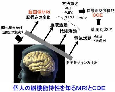
Today I am working on improving MRI and COE technology that extracts brain functions individually in our daily life through basic research and application, and train younger generations to create “brain culture” based on medicine and brain science (fig.4).
If we combine MRI and COE technology together, we can measure learning ability of a patient who may even appear to be in a vegetative state.
See my publications